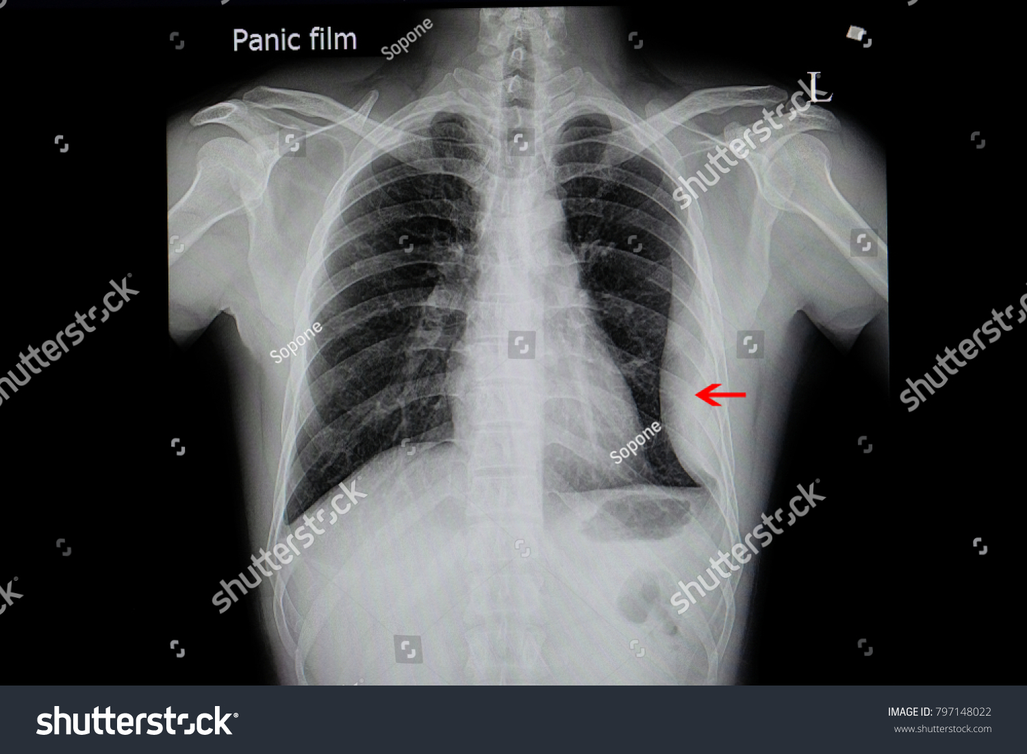
Loculated Pleural Effusion ~ Pleural Effusion In Major Fissure Ppt Powerpoint. Left pleural effusion with high density material at the posterior costophrenic angle. What are the different appearances of pleural effusion? Pleural effusions describe fluid between the two layer of tissue (pleura) that cover the lung and the lining of the chest wall. (vats) with lysis of adhesions is also a viable option for loculated effusions. The category 3 effusion meets at least one of the following criteria:
Pleural effusion occurs when fluid gets between the two layers of tissue that cover the. Or (3) the pleural fluid ph is less than 7.20 or the pleural fluid glucose is less than 60 mg/dl. Pleural effusion that is confined to one or more fixed pockets in the pleural space. Loculated pleural effusion must be included in the differential diagnosis of roentgenographic densities in the chest when seen in subcostal as well as in interlobar locations. Icu patients cannot sit up and the effusion layers posteriorly.
Pulmonology 16 years experience see below: The largest pocket of fluid is present posteriorly at the right lung base, with associated atelectasis and minor consolidation. This type of effusion is empyema unless proven otherwise. The pleural fluid is called a transudate if it permeates (transudes) into the pleural cavity through the walls of intact pulmonary vessels. Or (3) the pleural fluid ph is less than 7.20 or the pleural fluid glucose is less than 60 mg/dl.
A Loculated Pleural Effusion A Complex Pleural Effusion Is Shown With Download Scientific Diagram from www.researchgate.net February 09, 2021 a bilateral pleural effusion occurs when fluid between the lungs and chest dysfunctions, creating fluid accumulation. The pleura are thin membranes that line the lungs and the inside of the chest cavity and act to lubricate and facilitate breathing. If it is clear that there are multiple loculations then it is wise to avoid delay and proceed directly to this procedure. The pleural fluid is called a transudate if it permeates (transudes) into the pleural cavity through the walls of intact pulmonary vessels. Search for loculated pleural effusion. In vitro efficacy of varidase versus streptokinase or urokinase for liquefying thick purulent exudative material from loculated empyema. Loculated effusions, defined as effusions that do not shift freely in the pleural space, occur when there are adhesions between the visceral and parietal pleura. There is normally a small amount of fluid between these layers.
(2) the gram stain or culture is positive;
Indwelling pleural catheters (ipcs) are effective management options for malignant pleural effusion. The etiology of the pleural effusion determines other signs and symptoms. Pleural effusions in the intensive care setting. Pleural effusion is fluid buildup in the space between the layers of the pleura. Most malignant effusions can be controlled by thoracentesis and/or closed thoracostomy tube drainage and sclerosis of the pleural cavity. Many medical conditions can lead to it, so even though your pleural effusion may have to be drained, your doctor likely will target. Twenty of the 21 complicated parapneumonic effusions (including empyemas) showed loculation (95%). Tell a friend about us, add a link to this page, or visit the webmaster's page for free fun content. Encysted pleural fluid is visualized between the right upper and middle lobe (s). Of the 22 transudates, eight showed a loculated pleural effusion (36%) compared with 45 of 78 exudates (58%). In chf effusions are bilateral and more on right. If it is clear that there are multiple loculations then it is wise to avoid delay and proceed directly to this procedure. Icu patients cannot sit up and the effusion layers posteriorly.
Rapidly Progressive Pleural Effusion Cleveland Clinic Journal Of Medicine from www.ccjm.org A pleural effusion is an unusual amount of fluid around the lung. Search for loculated pleural effusion. The pleural fluid is called a transudate if it permeates (transudes) into the pleural cavity through the walls of intact pulmonary vessels. Encysted pleural fluid is visualized between the right upper and middle lobe (s). One layer rests directly on the lungs. Conventional chest radiography and computed tomography (ct) scanning are the primary imaging modalities that are used for evaluation of all types of pleural disease, but ultrasound and magnetic resonance. Symptomatic fluid loculation is a recognized complication of ipc use and is usually managed with intrapleural instillation of fibrinolytic drugs, such as tissue plasminogen activator (tpa). Or (3) the pleural fluid ph is less than 7.20 or the pleural fluid glucose is less than 60 mg/dl.
Pleural effusion is fluid buildup in the space between the layers of the pleura.
Symptomatic fluid loculation is a recognized complication of ipc use and is usually managed with intrapleural instillation of fibrinolytic drugs, such as tissue plasminogen activator (tpa). If it is clear that there are multiple loculations then it is wise to avoid delay and proceed directly to this procedure. Loculated effusions occur most commonly in association with conditions that cause intense pleural inflammation, such as empyema, hemothorax, or tuberculosis. Tell a friend about us, add a link to this page, or visit the webmaster's page for free fun content. The pleural fluid is called a transudate if it permeates (transudes) into the pleural cavity through the walls of intact pulmonary vessels. Pleural effusion is a rare complication and is generally similar to that seen in the rickettsial illnesses. The pleura is a thin piece of tissue with 2 layers. A pleural effusion is due to the manifestations of another illness. The space where the fluid is located is called the pleura, and it plays a vital role in the health. Normally, a small amount of fluid is present in the pleura. Pleural effusion occurs when fluid gets between the two layers of tissue that cover the. In chf effusions are bilateral and more on right. Encysted pleural fluid is visualized between the right upper and middle lobe (s).
Share :
Post a Comment
for "Loculated Pleural Effusion ~ Pleural Effusion In Major Fissure Ppt Powerpoint"


Post a Comment for "Loculated Pleural Effusion ~ Pleural Effusion In Major Fissure Ppt Powerpoint"